Friday, March 29, 2013

His first surgical repair - the Jatene Procedure


If you are here and still reading, thanks for being so patient!  Our family has been very busy over the last six months, living, moving and preparing for Alexander's surgery in Boston on April 24th.

Alexander’s first 7 days of life was spent in the Intensive Care Unit.  He underwent several diagnostic tests to better pinpoint his anatomy.  On the morning of his 8th day, we handed him helplessly to surgeons and hoped that he would survive.  For 115 minutes his tiny heart did not beat, his lungs did not breathe, his body was cold.  One-hundred fifteen minutes~nearly two hours~Alexander relied completely on heart-lung bypass to ensure his body and mind could survive the repair to his heart.

A newborn’s heart is about the size of a walnut.  Because of this minuscule size, our surgical team did not feel they could complete Alexander’s repair in one surgery, however, it was vital to get his ventricles pumping to their appropriate systems.

The ventricles (or lower chambers) are the muscles that pump the blood to where it needs to go.  They are structurally different as the right normally pumps just to the lungs, and the left pumps to the entire body.  The left ventricle is able to pump at high pressures for a very long time (a lifetime!) and the right pumps at a much lower pressure as the blood only needs to travel a short distance to the lungs.

With Alexander’s TGA (transposition of the great arteries) his left lung was pumping to his lungs and the right to his body.  Even though he had enough mixing of his oxygenated and deoxygenated blood to keep his tissues reasonably oxygenated, within just a few weeks, his left ventricle would start to atrophy, or weaken, as it was not pumping to his body.  In the meantime, his lungs would be getting more pressure and blood than they were intended and this would be bad for them as well.  If you can imagine the lungs like a balloon, but this balloon isn’t meant to be expanded endlessly, it is only meant to fill just to capacity and empty.  If it’s stretched too much or too often, it becomes weaker, just like a real balloon.


This procedure, while straightforward on paper, takes amazing precision as the great arteries are smaller than drinking straws and the tiny coronary arteries (vessels that supply oxygen-rich blood to the heart) are wire thin.  Once, while marveling with a nurse about the miracles these steady hands perform, she mentioned that many of the suture threads are so light that they float instead of hang.

While he was in the operating room, they also ligated his left-Superior Vena Cava (l-SVC), made repair to a Ventricular Septal Defect (VSD), and performed an atrial septoctomy.  
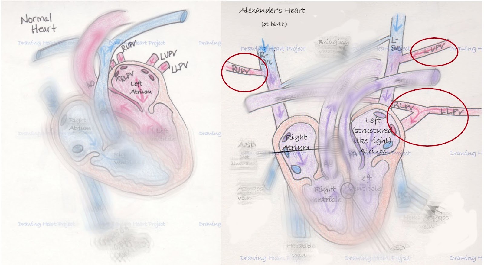
As I’ve described before, in normal anatomy one superior vena cava (SVC) drains the blood from the upper part of the body directly to the heart.  Alexander was born with one on each side of his body draining to each side of his heart.  He possesses a small bridging vessel between the two and surgeons elected to utilize that vein to promote better circulation.  Just below the bridging vessel, his l-SVC was tied off and severed, forcing the blood to cross the bridging vessel into the right SVC and into the right atrium.  This also left his Left Upper Pulmonary Vein (LUPV) to return blood back to the left side of his heart as it should.


Ventricular defects significantly affect the hemodynamics (blood flow patterns) inside the heart.  More or less blood flowing into the ventricles significantly change how well the muscle and valves perform.  To help mitigate the affect, doctors attempted to close a VSD in the heart wall.  The procedure was carried out well, but more defect remained for repair later.


Finally, surgeons performed an atrial septoctomy.  Because of his unique circulation, doctors felt that having the left and right atrium opened to each other to allow blood to mix more efficiently.  The surgeons removed the wall separating the left and right atria to create a large atrial septal defect (ASD) or a surgically common atrium.

Alexander emerged from surgery and was stabilized in the Cardiac Intensive Care Unit.  His chest was left open to allow for swelling and he remained intubated (as you can’t draw in breath independently while your sternum is split open.)  It was both frightening and reassuring to watch his tiny heart beat under a thin dressing.  His first hours were rocky, but not unexpected.  Four days later his chest was closed and two days following he was extubated.  This surgery staged us for his repair happening in April. 

After having a few complications in the first weeks, Alexander recovered well.  His current oxygen saturations are in the low 90s and he is just beginning to show us signs that his heart is holding him back.   We feel the timing of this surgery cannot be better.  He is strong, yet appearing to plateau.  Hopefully this means he'll have a wonderful recovery.

In the next few days, I plan on explaining the next repair Alexander needs for his special heart.


***I am not a nurse or doctor, nor have I received any formal medical training.  Any and all medical related information on Drawing Heart is the product of a mother’s desperation to understand and advocate for her child.  This blog is the culmination of countless hours of independent research and medical consultation and is meant only to communicate my understanding of Alexander’s condition.  It is not intended as medical advice.  As always, seek the advice of a qualified medical professional to explain your specific diagnosis.***


Monday, November 26, 2012

Intermission

It's been longer than I intended since I've made a new post.  As a family, we have been so busy and to add to that, last week we launched Our Baubles for Boston Campaign.  The response thus far has been tremendous, but it certainly keeps my fingers busy beading ornaments.  In the coming weeks, I hope to make a few posts regarding Alexander's surgical interventions, but for now just a quick update!


Sunday, October 7, 2012

Heterotaxy RAI vs LAI


(While reading this entry, please remember I am not a medical professional.  Also keep in mind that heterotaxy is as different as every patient.  Some individuals may have all the conditions described, or very few.  This is just a guide for basic understanding.)

I felt like I should bring in a special guest illustrator for this post.  Maybe I was a bit ambitious, but I enjoy giving others a chance, especially when the artist has life-long experience with heterotaxy.  At first he was eager, quickly sketching how the internal organs might look, he was very intense.


Then, he went all “diva” on me, demanded breastmilk, a cookie and a nap, whining something that sounded a lot like “I can’t possibly work under these deplorable conditions”.  Of course, I was forced to tuck 16 month old Alexander into his bed and go on without him, even though I think there may be some degree of accuracy to his unfinished drawing.

Humor aside, there are days when his scribbles make me think of heterotaxy.  The literal translation of heterotaxy is “different arrangement”.  There is not a consensus on exactly how that applies, but many feel that the heart is always involved (though, perhaps not to a great degree), and there is some abdominal ambiguity.

Looking at a person from the outside, it’s hard to imagine the complexity that lies on the inside.  Generally speaking, the outside appearance of a person is symmetrical between the left and right side.  A person’s arms are about the same size and length, their feet are about the same size, and the ears are near the same spot.  The left side is much like a mirror image of the right, or vise versa.

On the inside it’s the opposite.  The left side is completely different from the right.  The stomach is on the left side of the body, the liver on the right.  Even paired organs that have one organ on each side of the body are structurally different from each other.  For instance the left lung only has two lobes, while the right has three and the right adrenal gland is triangular shaped and the left horseshoe shaped.


Heterotaxy disrupts this asymmetrical balance.  During the early days of a pregnancy (between the 3rd and 6th week) the internal organs are forming.  They are created from tubes of tissue that are told how to twist and turn to form the intricate chambers of the heart, blood vessels and digestive tract.  In heterotaxy, these twists and turns don’t happen the way they should, and it’s not entirely understood why.  There are some genetic markers that indicate inherited heterotaxy, and sometimes the syndrome does run in families.  There is also some evidence that heterotaxy (whole or in part) could be related to Primary Ciliary Dyskensia (PCD) which affects the ability of tiny hairs (cilia) in our bodies to move things around they way they should.


Most cases of heterotaxy can be organized into two types.  There is Left Atrial Isomerism (LAI) and Right Atrial Isomerism (RAI).  To better understand what is happening, it helps to know the definition of isomerism.  Isomerism is a chemistry term that describes different compounds that are made up of the same parts (atoms), but they are bonded together differently, and that makes the compound completely different.  The use of isomerism when referring to heterotaxy describes optical isomerism, or when those compounds are arranged in such a way they are the mirror images of the other.

Remembering from earlier, think of the outside of our body as optically isomeric; it is made up of the same parts, but in a mirror image to the other.  However, the inside isn’t supposed to be, as there are drastic differences between the right and left internal organs. Therefore, in RAI, all the parts of the right side are present, but mirror imaged to the left, and in LAI all of the left-sided structures are there, but mirror imaged to the right.  This isn’t perfect, but it does help when knowing what to expect from a RAI patient versus a LAI.

In RAI, the structures of the heart are usually greatly affected.  Much of the heart lies on the left side of our bodies, so it makes sense that when the right side of the body is copied, the heart would suffer.  The lungs often end up as two tri-lobed lungs (as the right lung is tri-lobed), the stomach is on the right side of the body versus the left, the spleen is generally absent (asplenia).  The liver usually becomes transverse (midline) and the gall bladder can be affected, and the intestines are often malrotated and the kidneys and adrenal glands can suffer some anomalies.

In LAI one can almost infer the opposite to be true.  The heart tends to have less structural problems, but more prone to electrical ones as the heart’s natural pacemaker is located in the right atrium.  Often, the lungs are both bi-lobed (and bi-lobed lungs are slightly smaller than tri-lobed).  The liver can be midline and malrotated intestines might be present.  Many patients have polysplenia (or many spleens), and while one may think many is much better than none, often times they are not working.  This is known as functional asplenia.  Differences can also affect the kidneys and adrenals.

Because of how complex and extensive this syndrome is, every person affected is different.  One could talk to twenty patients and their families and hear twenty different stories and diagnoses.  These families have teams of doctors to help keep their children in the best possible health.  Our family has been fortunate that Alexander has been so well, but we’re also aware that the luck may not hold out forever.  Our hope is to continue to bring attention to this rare congenital syndrome.  Only through awareness can we convince people to care, and when enough people care change can happen.


I am not a nurse or doctor, nor have I received any formal medical training.  Any and all medical related information on Drawing Heart is the product of a mother’s desperation to understand and advocate for her child.  This blog is the culmination of countless hours of independent research and medical consultation and is meant only to communicate my understanding of Alexander’s condition.  It is not intended as medical advice.  As always, seek the advice of a qualified medical professional to explain your specific diagnosis.

Wednesday, September 19, 2012

Differences part 2


Because Alexander’s heart is complicated and I am wordy, I couldn’t quite keep the discussion about his heart in one post.  Drawing his heart, his anatomy, his condition has become very important to me.  I remember those first days when I was searching the web for “heterotaxy syndrome” and how little information would pop up.  I recall that most the articles were pitifully short and lacking details, or so in-depth I felt I needed a medical dictionary just to wade through the first paragraph.  Little more than a year later, that has changed for the better, and I want to continue the trend.  I want families to be able to understand Alexander’s diagnoses, and perhaps that would help them understand the ones they have been presented. 

Two Right Atria

The right atrium and the left atrium are structurally different.  They are intended to match with their corresponding ventricle.  For instance, the electrical impulses that tell the heart to beat originate in the right, and the left side of the heart is higher pressure system.  In Alexander’s heart, the atrium on the left side of his body is structured like a right atrium.  Many doctors say this shouldn’t cause a problem; if he had two left, he might need a pacemaker.  However, it is something of concern to me.  Alexander is unusual that he will have a two ventricle repair.  Not many heterotaxy-RAI patients are so lucky.  But what is luck for his ventricles, there really is a lack of information on how well his left-sided atrium will hold up.

Atrial Septal Defect (ASD) and Ventricular Septal Defect (VSD)

The separating wall between the left and right side of the heart is known as the septum.  It is solid and keeps the blue blood from mixing with the red.  In utero, there is a small hole (foramen ovale) between the left and right atrium, but like the ductus arteriosis (DA) it closes soon after birth.  In Alexander’s case, it was larger than normal and did not close on its own.  Alexander also had a moderate sized hole between his left and right ventricles.  Sometimes these defects close on their own, but Alexander’s were considered to be too large for that to happen.


Left-sided Inferior Vena Cava (IVC)

There have been many times when I have been baffled by Alexander’s anatomy, but none surprises me as much as this defect.  The IVC is a right-sided structure.  It brings all the deoxygenated blood back to the heart from the lower part of the body.  It forms from the two iliac veins (one from each leg) and travels just right of the spine to the right atrium.  All of the lower body’s veins flow into it as it climbs upward.  Before entering the atrium, the hepatic veins from the liver flow into the IVC.


Alexander’s IVC is much different.  He has a true left-IVC.  It is to the left of the Aorta and spine, and both of the iliac veins flow into it.  It continues past the renal (kidney) level and flows along what is referred to as a hemi-azygos continuation.  It then flows into the left side of the heart with no interruptions.  Alexander’s hepatic vein connects directly to the right atrium and his azygos vein connects to the right atrium instead of the SVC.  If you look at the vascular structure of Alexander’s heart with little regard for labels, you can see how much the left mirrors the right.  While this anomaly by itself may not cause issue, the flow from the IVC must be routed back to the proper side of the heart and any surgeon that may operate on him need to know of his abnormal vasculature to avoid mistakes.


Partial Anomalous Pulmonary Venous Return (PAPVR)

The pulmonary veins bring oxygen rich blood back to the heart to be delivered to the body.  Each pulmonary vein (there are typically four, but can be more) independently attaches to the left atrium.


In Alexander’s anatomy, none of his pulmonary veins attach like they typically should.  However, three of four veins do drain into the left atrium.  His right lower pulmonary vein (RLPV) and left lower pulmonary vein (LLPV) come together in what is known as a confluence before draining into the left atrium.  The left upper pulmonary vein (LUPV) drains into the L-SVC which drains to the left atrium.  The right upper pulmonary vein (RUPV) is the problematic one.  It drains into the R-SVC which drains into the right atrium.  In a heart with all of its flow being normal, this would reduce the oxygenated blood around 25%.  It also causes an increased workload on the right ventricle by adding additional blood volume.

Alexander arrived with a special kind of heart.  He has certainly had his cardiac team on their toes learning about his system.  But Alexander has other anomalies, too.  The effects of heterotaxy are far and wide on his tiny system, as is usual with those with this condition.  His body is like a tiny puzzle, every piece must be scrutinized and fit together exactly right to function well. 

I hope that in my next post I can describe heterotaxy on more of a broad scale, helping others understand the scope of this terrible condition.

***I am not a nurse or doctor, nor have I received any formal medical training.  Any and all medical related information on Drawing Heart is the product of a mother’s desperation to understand and advocate for her child.  This blog is the culmination of countless hours of independent research and medical consultation and is meant only to communicate my understanding of Alexander’s condition.  It is not intended as medical advice.  As always, seek the advice of a qualified medical professional to explain your specific diagnosis.***

Friday, September 14, 2012

Differences


Even with no knowledge of heart conditions, it’s easy to see Alexander’s heart is significantly different from a normal one.  It was created uniquely, just inside his tiny body and no where else.  However, it has and continues to work for him.  He’s a special heterotaxy-RAI guy that’s fortunate enough to have two ventricles.  The following is how I perceive his differences.  Please remember, I’m just a mom devouring medical documents, not a trained medical professional.

Dextrocardia (Incomplete)

Dextrocardia is when the heart is on the opposite side of the body.  Normally the human heart is located just on the left side of the body with the apex the furthest point left.  Oddly enough, the apex is not at the top of the heart, but the very bottom.  In complete dextrocardia, the heart is just on the right side, with the apex furthest right.  It would look something like this:

Alexander’s dextrocardia is incomplete.  His heart is completely midline, straight as you are looking at him, with the apex of his heart near the tip of his sternum.  


While in itself, this is not a problem, it has many effects.  It makes surgery more difficult, as the parts and structures surgeons are familiar with are not where they normally are, and creates new problems when trying to re-route heart plumbing.  It makes it more difficult to get accurate readings with ECG (electrocardiogram), it makes Echos (echocardiograms/ultrasound of the heart) harder to read.  Most of all, it’s indicative of other structural differences of the heart.

dextro-Transposition of the Great Arteries (d-TGA)

d-TGA generally happens when the heart is forming, near the 24th day of pregnancy.  Instead of twisting one last time, the Aorta and Pulmonary Artery remain parallel to one another.  While mother is carrying the baby, it has no effect, as the mother oxygenates the blood (baby doesn’t breathe) and the Aorta and Pulmonary Artery are connected by the Ductus Arteriosis (DA).  For such a simple problem, it devastates the circulatory system.  
  

At birth, a mother’s hormones stop being passed to baby and the DA begins to close.  The body gets less and less oxygen.   In d-TGA, instead of operating as an entire system, the heart operates as two closed loops.  The blue blood enters the heart on the right side and is immediately sent back out to the body through the Aorta.  On the left, blood rich in oxygen from the lungs enters the left and is sent directly back to the lungs.  The result is the body’s tissues are never oxygenated and the lungs receive blood at high pressure.   Without surgery, death is near certain by one year of age.

While Alexander was born with this condition, because of his other defects, d-TGA was not able to leave his body with no oxygen.

Dual Superior Vena Cava (SVC)

Because of Alexander’s heterotaxy, many structures that normally occur on the right are copied onto the left.  The SVC is the major vessel that drains all of the blood from the upper part of the body (from the heart up) back to the heart so it can be re-oxygenated.  It connects to the right atrium.  


Alexander also has one on his left side.  It connects into the left atrium much like the other side.  There was one small bridging vessel between the right and left SVCs.  The left SVC is problematic because it would return blue blood to the side of the heart that deals with red.  Instead of being able to head to the lungs and exchange its carbon dioxide for oxygen, it would simply mix with red blood and head out to the body.

Those are three of Alexander’s heart defects; he has five others.  Like all of his heterotaxy brothers and sisters, his anatomy is complicated.  In the interest of keeping blog sizes manageable, I’ll save those for later.


***I am not a nurse or doctor, nor have I received any formal medical training.  Any and all medical related information on Alexander’s Fight is the product of a mother’s desperation to understand and advocate for her child.  This series of notes is the culmination of countless hours of independent research and medical consultation and is meant only to communicate my understanding of Alexander’s condition.  It is not intended as medical advice.  As always, seek the advice of a qualified medical professional to explain your specific diagnosis.***

Sunday, September 9, 2012

A Normal Heart


When I began my journey understanding Alexander's heart, I realized that I must know what a regular heart does and looks like.  Only then, could I appreciate the special structures, the innovative compensation, and unique organ my son possesses in his chest.  It simply amazes me how a body can learn to deal with adversity and just how strong the body's instinct to survive is.

The heart is a miraculous machine.  It’s a pump that circulates blood throughout our bodies.  It delivers oxygen and nutrients to our tissues and disposes of the carbon dioxide and other waste.  It pumps approximately 60 times per minute, every minute of every day.  In a 50 year lifespan that’s approximately 1,576,800,000 times.  Yes, about one and a half BILLION times.

In a normal heart, blood enters from the body through veins on the right side.  Its oxygen stores are depleted and it’s blue.  It’s received in the right atrium, passes through the tricuspid valve into the right ventricle so it can head to the lungs.  When the right ventricle squeezes, it leaves through the pulmonary valve in the pulmonary artery.

The pulmonary artery branches and delivers blood to both lungs.  Here, the carbon dioxide waste is traded for oxygen and the blood is red again.  It returns to the heart through the pulmonary veins (there are usually 4 of these, but more can occur naturally) into the left atrium.  When the heart relaxes, the oxygenated blood passes through the bicuspid (or mitral) valve into the left ventricle.  With the next pump of the heart, the blood is pushed out of the aorta, through the aortic valve where it travels to the body to deliver its oxygen and nutrients.

This is a very elegant solution that supports every system in our body.  And every year 1 out of every 100 children born will have one that is less than perfect.  Congenital Heart Disease (CHD) claims more lives than all of childhood cancers combined and is the leading cause of death in children.  Our children need us to understand how this miraculous muscle works.

Alexander has no less than eight CHDs.  Less than 10 years ago, his diagnosis (Heterotaxy-Right Atrial Isomerism) had 80% mortality before the first birthday.  While the numbers are improving today, they still are frighteningly poor.  It is a constant worry, but odds we plan on defeating.
This is Alexander's heart beside a normal one.  It's obvious that it's different, but it's still strong.  His specific heart diagnoses are:

Dextrocardia (not complete)
d-TGA (dextro-Transposition of the Great Arteries)
Dual SVC
two right atria
ASD (Atrial Septal Defect)
VSD (Ventricular Septal Defect)
left-sided IVC
PAPVR (Partial Anomalous Pulmonary Venous Return)

Among children born with Heterotaxy-RAI, he is lucky.  His left ventricle is full size and fully functioning.  He lacked defects involving his pulmonary and aortic valves and it's hopeful that we can straighten out his circulatory system without compromising his quality of life.  We are hopeful and he is strong.


***I am not a nurse or doctor, nor have I received any formal medical training.  Any and all medical related information on Drawing Heart is the product of a mother’s desperation to understand and advocate for her child.  This blog is the culmination of countless hours of independent research and medical consultation and is meant only to communicate my understanding of Alexander’s condition.  It is not intended as medical advice.  As always, seek the advice of a qualified medical professional to explain your specific diagnosis.***

Saturday, September 8, 2012

Drawing Heart


***I am not a nurse or doctor, nor have I received any formal medical training.  Any and all medical related information on Drawing Heart Project is the product of a mother’s desperation to understand and advocate for her child.  This blog is the culmination of countless hours of independent research and medical consultation and is meant only to communicate my understanding of Alexander’s condition.  It is not intended as medical advice.  As always, seek the advice of a qualified medical professional to explain your specific diagnosis.***

 
From the moment Alexander was born he was making doctors ask questions.  Because of fetal imaging we knew he would be born with d-TGA (dextro-Transposition of the Great Arteries).  Traditionally, these infants have been called “blue babies” as the circulation did not allow oxygen into their blood and to their bodies.  Alexander was born pink and continued to be.  Doctors knew something else must be occurring for him to be so well oxygenated.

Within hours, Alexander was the main attraction in the NICU/CICU.  Every doctor came to look at him, tons of imaging was done, nurses were constantly puzzled about his true condition.  We had one doctor that consistently drew pictures for the other staff, trying to communicate Alexander’s entire heart status.  Terminology was constantly thrown out, and Matt and I weren’t nearly as educated on the topic as we are now.

At eight days old, they prepared to take him into surgery for the first time.  The cardiac surgeon talked to us about his conditions and the surgical plan when he spoke the words that inspired this project.

“He’s unique; no other like him.  You won’t find a picture of his heart in a book or online.  He’s as rare as they come.”

That upset me.  How could I, a “normal” parent, understand when the doctors are not only delivering diagnoses that I’ve never heard of, made up of multiple words that each need a google search, but have no real illustrations to go with it?  How could I grasp Alexander’s entire condition “blindly?”

The answer was a long amount of time, a ton of research and, eventually, sketching his anatomy.  It brought a great amount of confidence understanding his condition and being able to communicate it.  I’ve found myself sketching the same heart pictures over and over, refining them, explaining his special heart to the newest doctor.

That brings me to now, and the project that’s known as “Drawing Heart” on my hard disk.  I realized that if I had trouble understanding Alexander’s condition, others must have as well.  I decided that I would draw the structures and conditions that have become so familiar to me in hope that others would be able to better understand by looking and reading my own interpretations.  My goal is to raise awareness and understanding for all things heterotaxy.

For now I’ll just leave my drawing of Alexander’s special heart when he was born(so now a picture of it can be found online!), but I will explain it in a later post.  I hope “Drawing Heart” brings attention and increases understanding.